
- Life Sciences Division, Lawrence Berkeley National Laboratory, Berkeley, CA 94720 USA
- Department of Laboratory Medicine, University of California, San Francisco, CA 94143 USA
- Department of Radiation Oncology, New York University School of Medicine, New York, NY 10016 USA
Breast Cancer Research 2010, 12:R11 doi:10.1186/bcr2477
Received: 26 August 2009
Revisions requested: 21 October 2009
Revisions received: 20 January 2010
Accepted: 10 February 2010
Published: 10 February 2010
© 2010 Mukhopadhyay et al.; licensee BioMed Central Ltd.
This is an open access article distributed under the terms of the Creative Commons Attribution License (http://creativecommons.org/licenses/by/2.0), which permits unrestricted use, distribution, and reproduction in any medium, provided the original work is properly cited.
Most human mammary epithelial cells (HMEC) cultured from histologically normal breast tissues enter a senescent state termed stasis after 5 to 20 population doublings. These senescent cells display increased size, contain senescence associated β-galactosidase activity, and express cyclin-dependent kinase inhibitor, p16INK4A (CDKN2A; p16). However, HMEC grown in a serum-free medium, spontaneously yield, at low frequency, variant (v) HMEC that are capable of long-term growth and are susceptible to genomic instability. We investigated whether ionizing radiation, which increases breast cancer risk in women, affects the rate of vHMEC outgrowth.
Pre-stasis HMEC cultures were exposed to 5 to 200 cGy of sparsely (X- or γ-rays) or densely (1 GeV/amu 56Fe) ionizing radiation. Proliferation (bromodeoxyuridine incorporation), senescence (senescence-associated β-galactosidase activity), and p16 expression were assayed in subcultured irradiated or unirradiated populations four to six weeks following radiation exposure, when patches of vHMEC became apparent. Long-term growth potential and p16 promoter methylation in subsequent passages were also monitored. Agent-based modeling, incorporating a simple set of rules and underlying assumptions, was used to simulate vHMEC outgrowth and evaluate mechanistic hypotheses.
Cultures derived from irradiated cells contained significantly more vHMEC, lacking senescence associated β-galactosidase or p16 expression, than cultures derived from unirradiated cells. As expected, post-stasis vHMEC cultures derived from both unirradiated and irradiated cells exhibited more extensive methylation of the p16 gene than pre-stasis HMEC cultures. However, the extent of methylation of individual CpG sites in vHMEC samples did not correlate with passage number or treatment. Exposure to sparsely or densely ionizing radiation elicited similar increases in the numbers of vHMEC compared to unirradiated controls. Agent-based modeling indicated that radiation-induced premature senescence of normal HMEC most likely accelerated vHMEC outgrowth through alleviation of spatial constraints. Subsequent experiments using defined co-cultures of vHMEC and senescent cells supported this mechanism.
Our studies indicate that ionizing radiation can promote the outgrowth of epigenetically altered cells with pre-malignant potential.
Carcinogenic consequences of radiation exposure have historically been attributed to targeted effects - misrepaired DNA damage directly caused by dose-dependent ionization events in the cell of cancer origin. Radiation can also induce non-targeted effects - altered cytokines and signaling that affect the cellular composition and microenvironment of irradiated tissues(1), and non-mutational, but heritable changes that alter cell-cell interactions and induce persistent phenotypes associated with malignant progression(2)(3)(4). The potential carcinogenic contribution of these non-targeted effects, which are typically not linearly proportional to radiation dose, has not been well studied, particularly in primary human epithelial cells.
In this study, we used primary cultures of human mammary epithelial cells (HMEC) as an experimental system to directly evaluate the potential of ionizing radiation to promote the outgrowth of cells bearing a pre-malignancy-associated epigenetic change. In serum-free growth medium, HMEC from histologically normal breast tissues arrest growth after 5 to 20 population doublings, exhibit senescent morphologies, and express p16INK4A (CDKN2A; p16)(5)(6). This p16-dependent form of senescence, termed stasis, is distinguished by irreversible growth arrest with 2N DNA content (reviewed in(7)). Stasis is associated with poorly defined imbalances in signal transduction brought on by cell culture conditions or oncogene activation, but is not directly associated with DNA damage or dysfunctional telomeres(8)(9). Stasis requires activation of another well-known tumor suppressor, pRB, which functions downstream of p16, and serves as a block to indefinite proliferation (immortality) - a prerequisite for malignant transformation. The heterogeneous p16 expression observed in human breast epithelial cells in situ(10), and frequent aberrations in the p16-pRB pathway in human tumors(11), suggest that conditions that influence its expression and silencing have physiological and pathological relevance.
HMEC cultured in a serum-free medium spontaneously yield rare variant (vHMEC) cells in which p16 genes are methylated and silenced at frequencies that differ among normal specimens from women(12). In previous studies, such vHMEC have been shown to be susceptible to genomic instability associated with telomere and centrosome dysfunction(6)(13)(14). In some cases following carcinogen or oncogene exposure, these vHMEC give rise to immortalized clones bearing chromosomal aberrations commonly observed in primary human breast tumors(15). Here, we sought to determine whether low to moderate doses of radiation (5-200 cGy) would alter the rate of vHMEC outgrowth from non-malignant tissues. In replicate experiments using HMEC from four different specimens, we found that radiation caused shorter growth plateaus and significantly increased the rate at which p16(-) vHMEC grew out in long term cultures. Computer simulations using an agent-based model suggested that radiation accelerated selection of pre-existing vHMEC, a prediction that was confirmed experimentally. Thus non-targeted radiation effects can lower a critical cancer barrier by altering cell interactions to promote outgrowth of cells with pre-malignant phenotypes.
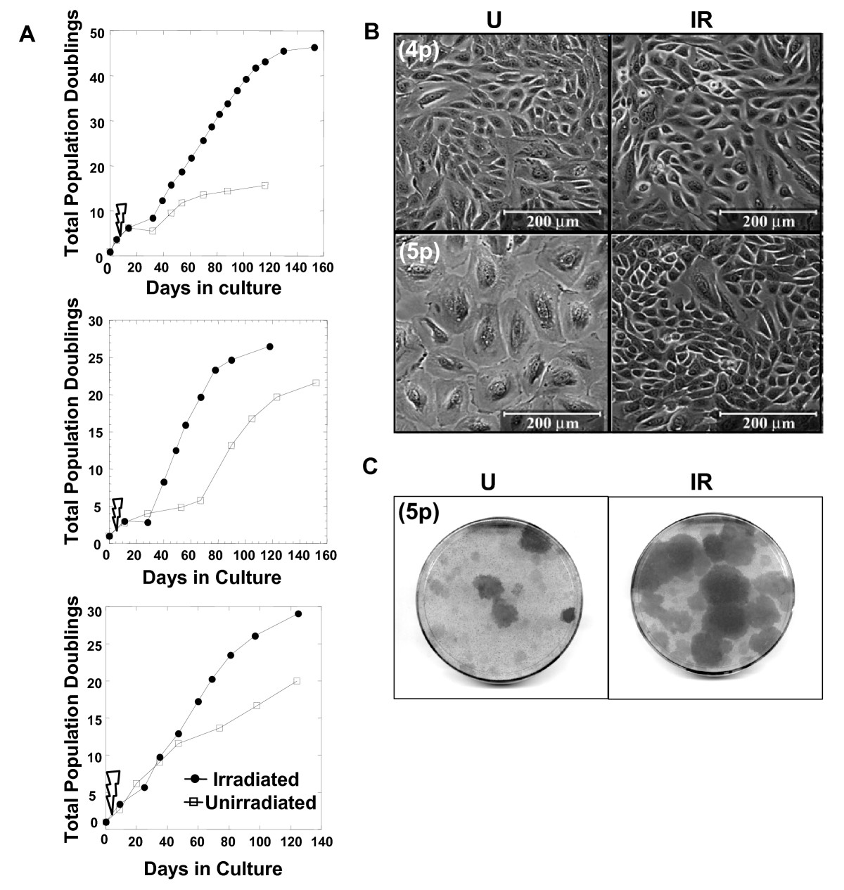
(A) Plots of the total population doublings of irradiated and unirradiated human mammary epithelial cells (HMEC) cultures versus days in culture for three experiments in which HMEC from different individuals were used (top to bottom; specimens B1450, B1400, and B1389). Each of the experimental points plotted in the graphs represents one passage. The time of irradiation between Passages 4 and 5 is indicated with a thunderbolt symbol. (B) Representative fields are seen in phase micrographs of unirradiated (U) and irradiated (IR) cultures at fourth passage (4p) 48 hrs after irradiation, or at fifth passage (5p) four to six weeks after irradiation. (C) Representative images of 5p plates stained with crystal violet illustrate the presence of larger and more frequent patches of actively growing cells in cultures derived from irradiated vs. unirradiated cells.
Click image to view larger.
Soluble factors induced by ionizing radiation, such as TGFβ, affect the growth of unirradiated cells(2). To determine whether the effect of radiation observed on the outgrowth of surviving cells was due to secreted factors, conditioned medium from irradiated or unirradiated cultures was added to the growth medium of unirradiated cultures. One set of irradiated and three sets of unirradiated B1400 or B1450 cultures were maintained separately. One unirradiated set served as a control and received only fresh medium at each feeding. A second unirradiated set received a 1:1 mixture of conditioned medium from irradiated cultures and fresh medium, while a third unirradiated set received a 1:1 mixture of conditioned medium from unirradiated cultures and fresh medium. Four replicates for each culture condition were maintained in this manner for four to six weeks. We did not observe any significant differences in the outgrowth of vHMEC in cultures supplemented with conditioned medium from irradiated or unirradiated cultures (data not shown). Thus soluble factors were not a likely cause of the differences observed.
We examined unirradiated and irradiated cultures 48 hrs following irradiation at 4p. The cultures exhibited similar cell densities and morphologies; areas of small proliferative cells were interspersed with large, flat non-mitotic cells in both (Figure 1B, upper panels). However, four to six weeks after irradiation, large uniform patches of small proliferative cells were evident earlier and were more numerous in the subcultured (5p) irradiated populations than in the unirradiated populations (Figure 1B, lower panels, and Figure 1C).
The expression of SA-βgal activity(17), a marker of cellular senescence, was measured in unirradiated and irradiated cultures when patches of vHMEC in unirradiated cultures reached 1.2 to 1.7 cm in diameter, typically four to six weeks post-irradiation. SA-βgal (-) cells were more abundant in irradiated than in the unirradiated cultures at these times (Figure 2A). To determine whether the differences observed between the percentages of SA-βgal (-) and (+) cells in irradiated and unirradiated cultures were statistically significant, 10 irradiated replicate cultures and 10 control replicate cultures from specimen B1450 were tracked independently. Flow cytometry was used to quantitate and compare the percentage of SA-βgal (-) cells in cultures four to six weeks following irradiation. At the time of harvest, 85.2 ± 1.6% of the cells in the irradiated cultures were SA-βgal (-), whereas 78.0 ± 2.6% of the cells in the unirradiated cultures were SA-βgal (-) (P < 0.001) (Figure 2B).

Representative fields are shown stained for (A) SA-βgal activity (blue reaction product) or (C) total p16 protein (green immunofluorescence). In (C), cell nuclei were counterstained with DAPI (blue). Inset = minus 1°Ab control. Note the greater presence of SA-βgal (-) and p16 (-) cells in the irradiated cultures. In (B) and (D), cytometry was used to quantitatively compare the percentages of positively and negatively staining cells in the corresponding cultures. In (B), flow cytometry results of an experiment performed using B1450 cultures are summarized in a mean ± SE scatter plot (P < 0.001, non-parametric Wilcoxon test). In (D), the percentages of p16 (-) cells in unirradiated and irradiated B1400 and B1450 cultures were determined using a Cellomics Array Scan VTI automated fluorescence microscopic imaging system. For each condition, staining was measured for 460 fields per well and summarized in a mean ± SE scatter plot (P < 0.0001, non-parametric Wilcoxon test).
Click image to view larger.
We next measured the expression of p16 protein by immunofluorescence in unirradiated and irradiated cultures harvested at identical times (Figure 2C). More than 20 random fields from unirradiated and irradiated B1450 and B1400 cultures were analyzed. Significantly more (70.5 ± 3.3% vs. 44.8 ± 3.5, P < 0.0001) cells were p16 (-) in the irradiated cultures than those in the unirradiated cultures (Figure 2D).

(A) Immunoblot analysis shows that p16 protein expression is very low in the eighth passage (8p) variant human mammary epithelial cells (vHMEC) cultures derived from both unirradiated and irradiated cells, but that it is readily induced to similar extents in both cultures treated with 5-aza-deoxycytidine (Aza). Lysates of HMEC undergoing stasis and MCF-7 cells were used as positive and negative controls, respectively. (B) p16 immunohistochemistry indicates that 8p vHMEC cultures derived from both unirradiated and irradiated cells contain primarily p16(-) cells, and that p16 protein can be induced in virtually all cells in both cultures by 72 hr treatment with 3.3 μM 5-aza-deoxycytidine. (C) DNA sequence of the CpG island region and a map of the -159 to +233 region of the p16 gene indicate the 35 CpG sites (CG or vertical lines) analyzed for methylation status, and their positions relative to the translational start site (ATG) and exon 1α. Primers used for PCR amplification are underlined. (D) The percent methylation of specific CpG sites (indicated by degree of shading) in the -159 to +233 region of the p16 gene was determined by sequencing of individual bisulfite-treated DNAs obtained from the indicated pre-stasis HMEC and vHMEC cultures from specimens B1450 and B1400.
Click image to view larger.
To determine whether radiation resulted in qualitative and/or quantitative differences in the methylation of CpG sites in the p16 promoter, we used bisulfite sequencing to investigate the methylation status of 35 CpG sites spanning from -159 to +233, relative to the translation start site (+1), in the core CpG island region of the p16 gene in two specimens, B1450 and B1400, with and without irradiation (Figure 3C). The majority of the CpG sites were unmethylated in pre-stasis 3p HMEC cultures harvested prior to stasis, whereas p16 promoter methylation was significantly increased in both unirradiated and irradiated vHMEC cultures, as indicated by the % methylation of individual CpG sites (Figure 3D). The methylation patterns were qualitatively and quantitatively distinct in irradiated and unirradiated cultures, however there was no discernable quantitative or qualitative correlation among p16 promoter methylation and passage level or radiation exposure status.
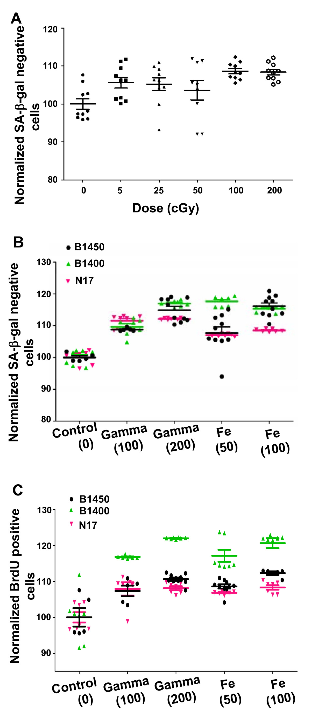
(A) Human mammary epithelial cells (HMEC) cultures (10 or more replicates per dose) from specimen B1450 were subjected to X-ray doses between 5 and 200 cGy. Each treatment group was normalized to a control group and the resulting means ± SE were plotted versus dose. (B and C) When assayed four to six weeks after irradiation with indicated doses (cGy) of 56Fe or γ-rays, indicated HMEC cultures showed statistically significant differences (P < 0.0001, non-parametric Wilcoxon test) in the percentages of (B) SA-βgal (-) and (C) BrdU (+) cells compared to control cultures. Scatter plots indicate the mean ± SE of between 5 and 10 independent replicates for three individual specimens assayed for each treatment.
Click image to view larger.
Densely ionizing 56Fe ions have been reported to be more potent inducers of genomic instability, cell transformation, and tumorigenesis than sparsely ionizing X-rays. The relative biological effectiveness (RBE) for cell killing of 1 GeV/amu 56Fe particles compared to X-rays was 1.80 at D10 (dose resulting in 10% survival by cell killing data) for HMEC(26). RBE values reported for other endpoints such as tumorigenesis in mice have been much higher(27)(28). We investigated the effect of radiation quality on the outgrowth of vHMEC. The progeny of cells generated from three breast tissue specimens (B1450, B1400, N17) were irradiated with densely ionizing 1 GeV/amu 56Fe particles and compared to those irradiated with sparsely ionizing 137Cs γ-rays four to six weeks after exposure, using flow cytometry to measure SA-βgal activity and BrdU incorporation (Figure 4B and 4C) in 5 to 10 replicates from each specimen for each treatment. Although the response of individual specimens to radiation exposure varied, as expected, the cultures irradiated with 137Cs γ-rays contained significantly more SA-βgal (-) and BrdU (+) cells, as in the X-ray experiments, than unirradiated controls (P < 0.0001; Wilcoxon test). Notably, the responses to 1 GeV/amu 56Fe ions were comparable to those of γ-rays and were not dose dependent.
ABMs are computer simulations that represent systems as collections of autonomous decision-making entities called agents(30). Each agent is programmed to respond to situations using a set of contextual rules. These models are non-deterministic, are typically based on multiple simulations so that a range of behaviors can be established for a population, and have proven to be very useful in predicting emerging properties from complex systems(31)(32)(33)(35)(36). Recently, ABMs have been used to predict the responses of cancer stem cell populations to ionizing radiation(37).
Based on experimental observations, we knew that the growth of HMEC is contact inhibited, that the area occupied by individual cells is reduced with increasing culture density, that all HMEC have limited replication capacity, that after a limited number of divisions most HMEC are capable of expressing p16 that initiates senescence, and that senescent cells do not readily replate following trypsinization. Thus we defined the following agent rules: 1) agents replicate as long as there is space surrounding them; 2) if there is no open space, agents can still replicate as smaller entities until reaching a minimum size; 3) when agents reach the maximum number of replications allowed, they stop dividing and their type changes permanently to a senescent type; 4) senescent agents do not reattach after detachment during subculture.
To validate these rules, we first collected experimental data describing growth characteristics of co-cultured HMEC and vHMEC imaged by time-lapse microscopy (note: vHMEC were obtained by taking HMEC that had escaped stasis in a previous experiment). A representative series of time-lapse images shown in Figure 5A illustrates how vHMEC outgrew labeled HMEC over time, as vHMEC could not express p16 and thus would not undergo stasis. Figure 5B shows the means and standard deviations of population doublings measured experimentally for each cell type in four replicate experiments. Note that the HMEC used here were at a higher passage than in Figure 1, as they rapidly underwent stasis on Day 4 after having divided on average only three times. In contrast, vHMEC cells grew until they reached confluence. Thus, to simulate this series of co-culture experiments, a maximum limit of three divisions for HMEC agents was set, whereas no division limit for vHMEC agents was set. As described above, Figure 5C illustrates the key rule for contact inhibition, showing how agents stop growing once they have reached a minimum size determined experimentally by measuring the average area of fully confluent cells. The corresponding predictions from the ABM using these limited assumptions correlated well with the experimental data (Figure 5B), validating our rules.
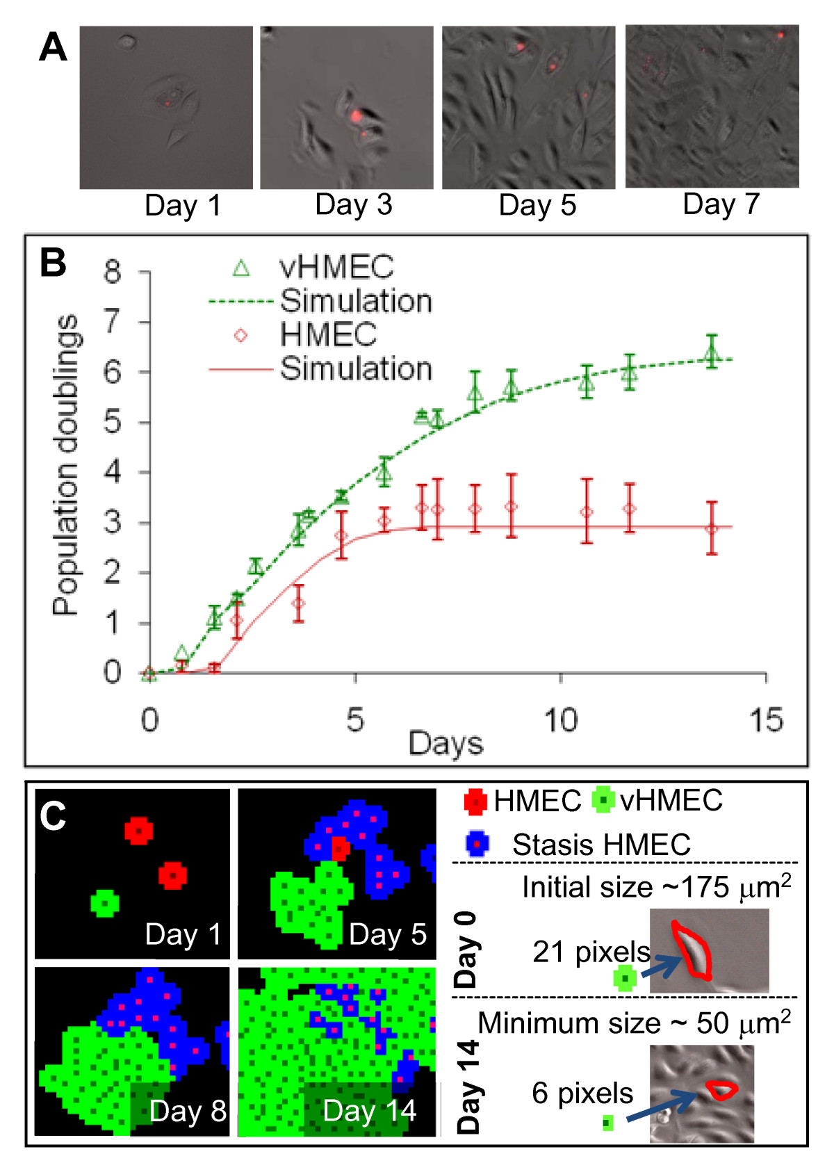
(A) Growth characteristics of co-cultured human mammary epithelial cells (HMEC) (labeled with vital dye Qtracker565; Invitrogen) and variant human mammary epithelial cells (vHMEC) (unlabeled) were recorded by time-lapse microscopy. An example is presented of the changes in proportions of cells present in a field of view monitored over seven days. (B) Growth of co-cultured HMEC and vHMEC was compared with ABM simulations. Each data point represents the mean of four biological replicates. Pre-stasis HMEC (red diamonds) stopped proliferating after four days in culture in contrast to vHMEC (green triangles) that continued to proliferate until the cultures achieved confluence. Solid lines show agent-based simulations, based on the initial cell densities, cell cycle duration during the proliferative phase (26 hours for both cell types), maximum cell compression observed at full confluence, and an intrinsic limit on the number of population doublings achievable by HMEC. Note that the simulations of the model were well correlated with the experimental data, indicating that kinetic growth rates could be predicted accurately for both cell types using a limited number of assumptions. (C) Illustration of growth simulation. Normal growing HMEC (red) undergo limited divisions before losing proliferative potential and undergoing stasis (blue). vHMEC (green) are capable of unlimited of divisions in this example. At low density (Day 1), most cells occupy a maximum area (that is, 21 pixels equivalent to 175 μm2, matching light microscopy measurements). Cells continue dividing as space becomes more restricted (Day 5), until the available adjacent space reaches a minimum of six pixels (50 μm2 at Day 14). Such simulations accurately recapitulate contact inhibition as measured by time-lapse microscopy.
Click image to view larger.
Agent-based modeling was then used to simulate the repetitive subculture experiments reported in Figure 1. Based on the growth curves reported in Figure 1A (in contrast to agents used in the validation step), a maximum of seven divisions for HMEC agents was set. We assumed that vHMEC were present initially at low but unknown frequencies in primary cultures and allowed a maximum number of 40 divisions for vHMEC agents. Using these assumptions and experimentally derived parameters, simulations led to growth curves very similar to those observed in Figure 1A, with the same significant population doublings' plateaus observed when the majority of the cells entered stasis (that is, after seven population doublings when assuming that 0.25% of initial agents were vHMEC agents, as illustrated in Figure 6A and 6B). The plateau widths reflected the time taken by vHMEC agents to overgrow the senescent HMEC populations, and were inversely proportional to the fraction of vHMEC agents present in the initial populations; a relationship that could be fitted accurately by an inverse power function (Figure 6C). Assuming that the population doublings' plateau indicated the period of selection of a minority of vHMEC present in primary cultures, the model could then inform us of the initial proportions of vHMEC in real specimens by extrapolating the plateau widths measured in these specimens with the fit obtained in Figure 6C (that is, for the three unirradiated cultures profiled in Figure 1A, plateau durations of 23, 60, and 10 days corresponded to initial vHMEC proportions of 0.25, 0.06, and 3.5%, respectively). Anecdotally, we found that the initial percentage of vHMEC present in the primary culture derived from a reduction mammoplasty specimen (B1400) was similar to values found for reduction mammoplasties in an earlier report(29), but that the percentages of vHMEC in primary cultures derived from prophylactic mastectomies (B1450, B1389) were higher. Data from additional specimens of each type will be needed to determine whether this is a statistically significant correlation.

(A and B) In a model extending over several passages, cells are initially plated at 20% confluence and replated when the populations reach 80% confluence. By Passage 3, most human mammary epithelial cells (HMEC) have undergone seven divisions, and enter stasis. As a result, the time to confluence is extended, and a population doubling plateau is observed. Ultimately, the variant human mammary epithelial cells (vHMEC) take up the available area, and exponential growth resumes. (C) Changing the initial density of vHMEC leads to variable plateau periods. This dependence is fitted by a power function that allows the calculation of the initial density of vHMEC present in the primary culture based on plateau length. The results of individual simulations are plotted as points.
Click image to view larger.
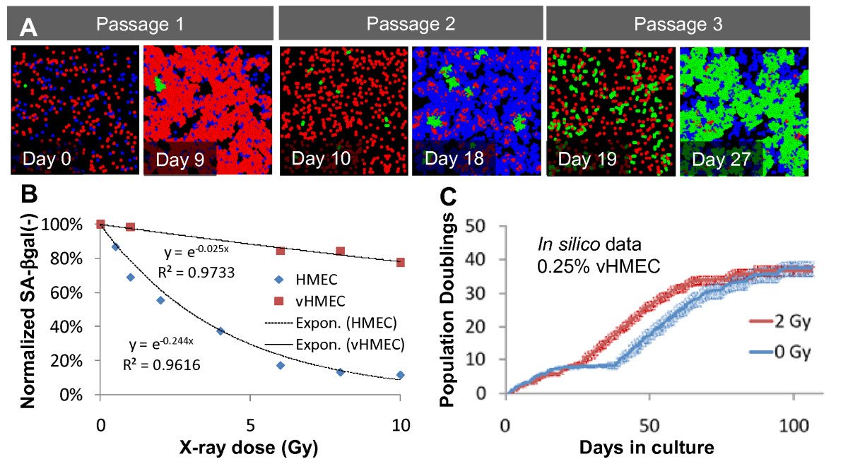
(A) Time snapshots of one representative simulation of sequential subcultures executed using the same initial conditions as in Figure 6A, but including prematurely senescent human mammary epithelial cells (HMEC) agents (labeled blue) induced by exposure to 2 Gy of X-rays. Note the earlier and more robust outgrowth of vHMEC agents (labeled green) in this simulation versus the simulation shown in Figure 6A. (B) Growing pre- and post-stasis HMEC from the same individual were exposed to the indicated doses of X-rays, then stained and evaluated for SA-βgal expression. The resulting data were then normalized to the percentage of SA-βgal(-) HMEC in the respective unirradiated populations. Fitted curves were of the form (e(-α.D)) where D is the dose and α the fitted coefficient; R2 values were >0.96 in both cases. These fitted curves were used to model radiation-induced senescence in the simulation depicted in (A). (C) Agent-based modeling was used to simulate the growth kinetics of HMEC cultures exposed to 0 or 2 Gy of X-rays. Data ± SE for five independent simulations for each condition are plotted. Note that the model predicts a shorter growth plateau in the irradiated sample, in accord with experimental observations.
Click image to view larger.
A major challenge is to understand how cellular responses to radiation are integrated in a multicellular context to affect human health. In this study, we show how radiation can promote the outgrowth of pre-malignant cells by accelerating senescence of normal cells. We used ABM to show that the rapidity with which radiation-resistant vHMEC selectively populate cultures can be predicted by the initial proportion of the vHMEC present in primary cultures, the differential rate at which sensitive HMEC succumb to radiation-induced loss of growth potential, and the physical space available for expansion.
Together with p53, the p16 protein functions as a sentinel that integrates various cellular signals and stresses to limit proliferation. Methylation of the p16 gene has been identified in situ in histologically normal mammary epithelial cells of disease-free women(29). A Luria-Delbrück fluctuation analysis suggests that such cells may be the source of vHMEC that continue to proliferate after most HMEC in long term cultures have undergone p16-associated stasis(29). Expression of p16 appears to be strongly selected against in many human solid tumors, including those of the breast, where the gene encoding p16 is deleted in approximately 20%(38) and inactivated epigenetically in an additional 20%(39) of cases. Interestingly, exposure of workers in nuclear weapons manufacturing facilities to plutonium, for example, has been strongly linked (P = 0.03) to methylation of the p16 gene in lung adenocarcinomas(40). Renal cell carcinomas from patients living in areas contaminated by the Chernobyl accident have also shown aberrant hypermethylation of the p16/p14 locus(41).
As we and others have observed, radiation itself does not appear to be a direct inducer of p16 (Additional file 1). Our new results indicate that radiation-induced stress can be integrated with p16-inducing factors that cause premature growth arrest and senescence. ABM simulations showed that radiation can advance the selection process, allowing vHMEC to overtake and fill the voids created by the prematurely senescing normal cells. A similar model, in which lesion growth is driven by opportunistic expansion of apoptosis-resistant p53 mutant cells, has recently been proposed for UVB-induced squamous cell carcinomas(42). The expansion of such a variant population in situ would be expected to expand the target size in which additional malignancy promoting aberrations could occur, especially since this population has been shown to be particularly susceptible to genomic instability caused by telomere dysfunction and centrosome irregularities(6)(13)(14).
p16 immunohistochemistry indicates that 3p HMEC cultures derived from specimen N17 did not express detectable p16 protein 24 hrs after irradiation with 2 Gy X-rays. Indicated negative and positive controls for specific antibody-dependent staining are shown in left panels.
Format: PDF Size: 85KB Download file
This file can be viewed with: Adobe Acrobat Reader
While accurately modeling the accelerated outgrowth of vHMEC from irradiated cultures, the current ABM does not explain why, in some cases, the total number of population doublings achieved by vHMEC from irradiated cultures exceeded that of vHMEC from unirradiated controls. This outcome could be due to either a radiation-induced increase in the number of vHMEC or modification of the phenotype of the existing vHMEC. Since the phenotypic feature of the vHMEC that allows them to avoid or overcome stasis is the stable repression of p16 expression, one possibility is that radiation directly affects epigenetic process(es) involved in the initiation or maintenance of this repression. DNA methylation-associated silencing of mammalian genes has been proposed to be a process, during which instances of spontaneous gene reactivation are initially frequent, becoming progressively less frequent over the course of multiple cell divisions (43)(44)43,44]]. In agreement with this hypothesis, recent experiments indicate that p16 gene methylation occurs progressively in clonal vHMEC populations after silencing has occurred(45). Irradiation may lead to acceleration of this process and thus reduce the frequency of spontaneous p16 reactivation. Indeed, while occasional p16(+) cells could be observed in 8p post-stasis cultures, they were more frequent in the progeny of unirradiated versus irradiated cultures (data not shown). The mechanism responsible for this difference remains to be discovered, but may involve radiation effects on sentinel proteins such as p53. Notably, radiation has been reported to relieve p53-mediated repression of DNA methyltransferase 1 (Dnmt1) in human colon carcinoma cells(46). Under certain circumstances, Dnmt1, which has been shown to be continually required for p16 repression(47), will catalyze de novo methylation of specific promoter CpG islands(48). Thus, radiation, acting through transient activation of p53, could cause de novo methylation and more stable silencing of the p16 gene.
We have found that radiation can indirectly promote the outgrowth of a putative pre-malignant breast cell population by accelerating senescence of normal breast cells. Differential sensitivity of these cell populations to radiation-induced senescence has not previously been demonstrated. While the mechanism responsible for this differential sensitivity is beyond the scope of our present investigation, the work advances a systems-based paradigm of carcinogenic activity by a prototypic genotoxic agent, ionizing radiation. The ABM simulation of well-described HMEC phenotypes in primary cultures helped to distinguish between induced vHMEC and altered population kinetics as plausible explanations for the observed response to radiation. ABM showed that differential sensitivity to radiation-induced senescence can at least partly explain the enhanced outgrowth of vHMEC from irradiated cultures. This simple in vitro system illustrates the concept that heterogeneous responses of individual cells within a population must be integrated to achieve a system-level understanding of radiation effects.
ABM: agent-based model; FBS: fetal bovine serum; FDG: fluorescein digalactoside; HMEC: human mammary epithelial cells; LET: linear energy transfer; p: passage; p16: p16INK4A; PBS: phosphate buffer saline; RBE: relative biological effectiveness; SA-βgal: senescence associated β-galactosidase; v: variant.
The authors declare that they have no competing interests.
RM performed cell culture, molecular and cellular assays, participated in the design, analyzed and interpreted data, and helped to draft the manuscript. SC designed and performed imaging assays and agent-based modeling, analyzed and interpreted data, and helped to draft the manuscript. AB and WCH acquired tissue samples and established primary HMEC cultures, and WCH assisted with imaging assays. MHBH and PY initiated the studies, participated in the design and coordination of the study, helped analyze and interpret data, and helped to draft the manuscript. All authors have read and approved the manuscript.
This work was funded by a NASA Specialized Center of Research, a Breast Cancer and the Environment Research Center Grant U01 ES012801 from the National Institute of Environment Health Science (NIEHS) and the National Cancer Institute (NCI), and the Office of Health and Environmental Research, Health Effects Division, United States Department of Energy (contract no. DE-AC02-05CH11231). A.V. Bazarov was supported by Flight Attendant Medical Research Institute Young Clinical Investigator Award No. 032122 and Komen for the Cure Research Grant No. BCTR0707231 and W.C. Hines received support from Komen for the Cure postdoctoral fellowship PDF0707408.
